The cardiac cycle is controlled by a portion of the autonomic nervous system (that part of the nervous system which does not require the brain's involvement in order to function). As we will see in the next chapter, nerve cells maintain a membrane polarization (more negatively charged ions inside the cell than outside) in their resting state. When the nerve cell "fires", the membrane depolarizes and the nerve impulse is transmitted to the next cell. One by one, but very quickly, the nerve impulse is "propagated" to the muscles which perform the required action.
The first step in the cardiac cycle is atrial depolarization. During this step, the atrium (which is filled with blood coming in from the circulatory system) contracts, pushing blood into the ventricle to fill it. The second step is ventricular depolarization. Here, the ventricle contracts (systole), pushing blood into the aorta, and the atrium is repolarized. Finally, the ventricles repolarize, during which time the atrium relaxes and refills. There are actually two atria and two ventricles in the heart; the left pair take oxygenated blood from the pulmonary subsystem and pump it into the rest of the body. The right pair take deoxygenated blood from the body and pump it to the lungs. We will ignore the latter pair for simplicity.
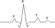
By means of an electrocardiogram (EKG), the successive depolarizations can be recorded. They are traditionally notated as follows:
The P site (often called the "P wave") represents the depolarization of the atria (both pairs function simultaneously). The R site (center of the "QRS complex") represents ventricular depolarization. The T site ("T wave") represents ventricular repolarization. The P and R waves precede actual contraction by a short time interval. The "U wave" is not usually present; it could represent repolarization of the "Purkinje fibers" at the "far end" of the ventricular nerves.
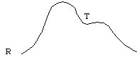
The sounds made by the heart during the cardiac cycle are observed at the S and T waves. The blood pressure in the aorta rises from the R site until just preceding the T wave, falls slightly and rises again at the end of the T wave, and then falls continuously until the next R wave:
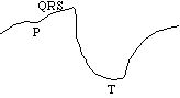
The ventricular volume varies as follows:
Disrythmias are caused by disturbances in automaticity (rate variations or premature depolarizations) and conductivity (blocked nerve impulses). Several types of disrythmia are:
The Mathematica examples show the blood pressure variation for many of these dirythmias, and may be useful in the next section.
The next section is about blood velocity and turbulence.
If you have stumbled on this page, and the equations look funny (or you just want to know where you are!), see the College Physics for Students of Biology and Chemistry home page.
©1996, Kenneth R. Koehler. All Rights Reserved. This document may be freely reproduced provided that this copyright notice is included.
Please send comments or suggestions to the author.